Friday 31 August 2018

Why does adenine pair with uracil in RNA ?

In RNA, uracil base-pairs with adenine and replaces thymine during DNA transcription. Methylation of uracil produces thymine.[10] In DNA, the evolutionary substitution of thymine for uracil may have increased DNA stability and improved the efficiency of DNA replication (discussed below). Uracil pairs with adenine through hydrogen bonding. When base pairing with adenine, uracil acts as both a hydrogen bond acceptor and a hydrogen bond donor. In RNA, uracil binds with a ribose sugar to form the ribonucleosideuridine. When a phosphate attaches to uridine, uridine 5'-monophosphate is produced.[6]
Uracil undergoes amide-imidic acid tautomeric shifts because any nuclear instability the molecule may have from the lack of formal aromaticity is compensated by the cyclic-amidic stability.[5] The amide tautomer is referred to as the lactamstructure, while the imidic acid tautomer is referred to as the lactim structure. These tautomeric forms are predominant at pH 7. The lactam structure is the most common form of uracil.
Uracil tautomersAmide or lactamstructure (left) and imide or lactimstructure (rightracil also recycles itself to form nucleotides by undergoing a series of phosphoribosyltransferase reactions.[2]Degradation of uracil produces the substrates aspartate, carbon dioxide, and ammonia.[2]
 → H3NCH2CH2COO + NH4+ + CO2
Oxidative degradation of uracil produces urea and maleic acid in the presence of H2O2 and Fe2+ or in the presence of diatomic oxygenand Fe2+.
Uracil is a weak acid. The first site of ionization of uracil is not known.[11] The negative charge is placed on the oxygen anion and produces a pKa of less than or equal to 12. The basic pKa = -3.4, while the acidic pKa = 9.389. In the gas phase, uracil has 4 sites that are more acidic than water.[12] a

Protein synthesis

 genetic information stored in DNA molecules is used as a blueprint for making proteins. Why proteins? Because these macromolecules have diverse primary, secondary and tertiary structures that equip them to carry out the numerous functions necessary to maintain a living organism. As noted in the protein chapter, these functions include:
• Structural integrity (hair, horn, eye lenses etc.). 
• Molecular recognition and signaling (antibodies and hormones). 
• Catalysis of reactions (enzymes).. 
• Molecular transport (hemoglobin transports oxygen). 
• Movement (pumps and motors).
The critical importance of proteins in life processes is demonstrated by numerous genetic diseases, in which small modifications in primary structure produce debilitating and often disastrous consequences. Such genetic diseases include Tay-Sachs, phenylketonuria (PKU), sickel cell anemia, achondroplasia, and Parkinson disease. The unavoidable conclusion is that proteins are of central importance in living cells, and that proteins must therefore be continuously prepared with high structural fidelity by appropriate cellular chemistry.
Early geneticists identified genes as hereditary units that determined the appearance and / or function of an organism (i.e. its phenotype). We now define genes as sequences of DNA that occupy specific locations on a chromosome. The original proposal that each gene controlled the formation of a single enzyme has since been modified as: one gene = one polypeptide. The intriguing question of how the information encoded in DNA is converted to the actual construction of a specific polypeptide has been the subject of numerous studies, which have created the modern field of Molecular Biology.

Repair of DNA damaging

One of the benefits of the double stranded DNA structure is that it lends itself to repair, when structural damage or replication errors occur. Several kinds of chemical change may cause damage to DNA:

• Spontaneous hydrolysis of a nucleoside removes the heterocyclic base component.
• Spontaneous hydrolysis of cytosine changes it to a uracil.
• Various toxic metabolites may oxidize or methylate heterocyclic base components.
• Ultraviolet light may dimerize adjacent cytosine or thymine bases.

All these transformations disrupt base pairing at the site of the change, and this produces a structural deformation in the double helix.. Inspection-repair enzymes detect such deformations, and use the undamaged nucleotide at that site as a template for replacing the damaged unit. These repairs reduce errors in DNA structure from about one in ten million to one per trillion

A different nucleic acid -RNA

The high molecular weight nucleic acid, DNA, is found chiefly in the nuclei of complex cells, known as eucaryotic cells, or in the nucleoid regions of procaryotic cells, such as bacteria. It is often associated with proteins that help to pack it in a usable fashion. 
In contrast, a lower molecular weight, but much more abundant nucleic acid, RNA, is distributed throughout the cell, most commonly in small numerous organelles called ribosomes. Three kinds of RNA are identified, the largest subgroup (85 to 90%) being ribosomal RNA, rRNA, the major component of ribosomes, together with proteins. The size of rRNA molecules varies, but is generally less than a thousandth the size of DNA. The other forms of RNA are messenger RNA , mRNA, and transfer RNA , tRNA. Both have a more transient existence and are smaller than rRNA. 
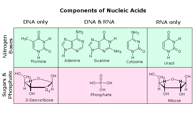
All these RNA's have similar constitutions, and differ from DNA in two important respects. As shown in the following diagram, the sugar component of RNA is ribose, and the pyrimidine base uracil replaces the thymine base of DNA. The RNA's play a vital role in the transfer of information (transcription) from the DNA library to the protein factories called ribosomes, and in the interpretation of that information (translation) for the synthesis of specific polypeptides. These functions will be described later.
A complete structural representation of a segment of the RNA polymer formed from 5'-nucleotides may be viewed by clicking on the above diagram

Chemical nature of DNA

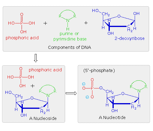
The polymeric structure of DNA may be described in terms of monomeric units of increasing complexity. In the top shaded box of the following illustration, the three relatively simple components mentioned earlier are shown. Below that on the left , formulas for phosphoric acid and a nucleoside are drawn. Condensation polymerization of these leads to the DNA formulation outlined above. Finally, a 5'- monophosphate ester, called a nucleotide may be drawn as a single monomer unit, shown in the shaded box to the right. Since a monophosphate ester of this kind is a strong acid (pKa of 1.0), it will be fully ionized at the usual physiological pH (ca.7.4). Names for these DNA components are given in the table to the right of the diagram. Isomeric 3'-monophospate nucleotides are also known, and both isomers are found in cells. They may be obtained by selective hydrolysis of DNA through the action of nuclease enzymes. Anhydride-like di- and tri-phosphate nucleotides have been identified as important energy carriers in biochemical reactions, the most common being ATP (adenosine 5'-triphosphate).
 

Names of DNA Base Derivatives

Base
Nucleoside
5'-Nucleotide
Adenine2'-Deoxyadenosine2'-Deoxyadenosine-5'-monophosphate
Cytosine2'-Deoxycytidine2'-Deoxycytidine-5'-monophosphate
Guanine2'-Deoxyguanosine2'-Deoxyguanosine-5'-monophosphate
Thymine2'-Deoxythymidine2'-Deoxythymidine-5'-monophosphate
A complete structural representation of a segment of the DNA polymer formed from 5'-nucleotides may be viewed by clicking on the above diagram. Several important characteristics of this formula should be noted.
• First, the remaining P-OH function is quite acidic and is completely ionized in biological systems.
• Second, the polymer chain is structurally directed. One end (5') is different from the other (3').
• Third, although this appears to be a relatively simple polymer, the possible permutations of the four nucleosides in the chain become very large as the chain lengthens.
• Fourth, the DNA polymer is much larger than originally believed. Molecular weights for the DNA from multicellular organisms are commonly 109 or greater.

What are secondary mediators ?

Secondary mediators are produced after target cell activation or released by the break down of phospholipids membrane during the process of degarnulation. Some of the secondary mediators are leukotrienes, various cytokines, prostaglandins etc

What is Atopic dermatitis?

Atopic dermatitis is an inflammatory skin disease. This disease is observed frequently in young children. There will be skin eruptions

What is erythroblastosis fetalis ?

It is a hemolytic disease, which develops in newborn. Maternal IgG antibodies cross the placenta and destroy the red bleed cells. This develops when an Rh+ expresses an Rh antigen on blood cells that the mother does not express.

What is the Rhogam ?

Is an antibody that binds to any of the blood cells, enter the mother’s blood circulation, and facilitate their clearance by activation of B-cells and memory cell production

Sunday 26 August 2018

What is size exclusion chromatography ?

In size exclusion, The sample is loaded in a column having small gel beads. The gel beads can be of Cellulose, Agarose, Silica etc. The beads are designed such that it has small pores of varying size (depending on the sample you want to separate). The smaller proteins/molecules enter the beads easily and thus have to migrate larger distance. On the counterpart, the larger proteins/molecules will not interact with the beads as the size of pores of the beads is too small for them to enter. Thus, the larger proteins/molecules have to migrate less distance and eluted first through the column.
The large molecules pass through the spaces present outside the beads. The Volume which is not occupied by the beads is called Void Volume and denoted by Vo. The Volume present inside the beads (as the beads are porous) is called Included Volume and denoted by Vi. The volume inside the bead which is occupied by the protein/molecules inside the bead is denoted by Mi.
Mi = Kd * Vi * C.
Where Kd = Partition Coefficient and C = Concentration.
Image[1]
In case of Gel electrophoresis, the larger molecules cannot migrate faster. It is because the pore size of the gel is small and the molecules have to migrate through the pore. There is not void volume in this case from where the molecules can easily migrate. Thus, the molecules have to migrate through the pores. This implies that larger the molecules, more will be the retardation force/friction experienced by those molecules. Hence, the migration of smaller molecules will be easier than the larger molecules as they encounter the least resistance during the movement in the gel.

What is affinity chromatography ?

Affinity chromatography technique exploits the property of the biomolecules such as enzymes to bind specifically to the substrate or any functional group[1].
Examples:
  1. Enzyme binding to the substrate.
  2. Antibody with specific Antigen.
  3. Hormone binding to receptor.
Requirements:
  1. Ligands which has an affinity for the substrate.
  2. Matrix to hold the ligand.
  3. The substrate which is your molecule of interest.
  4. Allosteric Binding site.
  5. Single step Purification.
  6. Spacer arms so as to create space between the ligand and the matrix. If the ligand and matrix are close to each other, then due to steric hindrance, the molecule of interest would not be able to bind the allosteric binding site on the ligand.
Properties of Matrix:
  • The matrix should be inert and stable. Its property should not change due to change in pH, Ionic strength and temperature during the elution technique.
  • The matrix should have multiple binding sites for the Ligands. Thus, more the ligand binds the matrix, greater the resolution.
  • The matrix should be porous so that surface area increase.
  • Example of matrices are sepharose, agarose, etc.
Properties of Ligands:
  • The ligand should have good affinity and specificity for the molecules of interest.
  • The ligand should bind the molecule of interest in reversible fashion. If the molecule binds to the ligand with a very high affinity, then the molecules will not be eluted out.
  • The ligand should have two binding sites. One to bind with the matrix and the other to bind with the molecule of interest.
  • The two binding sites should not overlap. If so, then spacer arms should be used to attach matrix and ligand. Spacer arms such as Diaminopropanol are commonly used[2].
  • Examples of ligands: Heparin ligands are used to isolate the fibronectins.

What is thin layer chromatography ?

Thin layer chromatography is a planar chromatographic technique which is used to separate and analyse the non-volatile compounds. It is simple and moderately sensitive. It is performed on a glass plate or thin foil made up of aluminium or plastic on which desired adsorbent is finely layered. Adsorbents like silica, aluminium oxide, or cellulose are commonly used in TLC.
TLC can also be modified for separation of desired components. The modification includes applying the principle of partition chromatography, Ion-exchange chromatography and Gel filtration (Size-exclusion) chromatography[1][2][3].
Types: Ascending and Descending Thin Layer Chromatography.
Stationary Phase: Silica, Alumina, Keiselguhr, Magnesium silicate etc.
Mobile phase: Polar solvents (Water, Methanol, Ethanol) and Non-Polar solvents (Hexane, Trichloroethylene, carbon tetrachloride).
Rf is also called as Retention factor or Retardation factor. It is the ratio of distance travelled by the sample to the distance travelled by the solvent.

What is paper chromatography

Paper chromatography is an analytical technique used to separate mixtures into constituent components.
Paper chromatography principle: It consists of three components – stationary phase, mobile phase and the mixture to be separated. The mixture is allowed to move on the stationary phase using the mobile phase due to capillary action. Due to differential interaction of the different components of the mixture with the stationary phase, they move at different speeds and thus, get separated.
Steps followed in this paper chromatography experiment have been listed below:
• Take a filter paper strip (stationary phase) of 10 cm and make marks on both ends.
• Put a drop of the blue ink (analyte) using glass capillary or splinter near one of the marks on the filter paper strip.
• Take some tap water (mobile phase) in a test tube.
• Put the chromatography strip in the test tube. Make sure that the sample spot is above the water level.
• Wait till the water along with the sample reaches the top marking. Remove the strip from test tube to view the results.
Different components of blue fountain ink are now separated. Filter paper strip is made of cellulose, which is a polymer of glucose, which in turn consists of many polar groups. The analyte we used has different components which have different polarities and thus they interact differently with the stationary phase. Due to this difference in polarities, individual components of the analyte move up the filter paper, along with water, at different speeds. More polar the compound, stronger is the interaction with the stationary phase and hence slower is its speed. On the contrary, less polar the compound, weaker is its interaction with the stationary phase and hence faster the movement along the filter paper.
Paper chromatography applications: Paper chromatography has a wide range of applications such as in separation of amino acids, biochemicals in urine and also for determination of hormones, drugs etc. in the pharma sector.

What happens when man drinks too much?

When we drink, alcohol goes to our stomach and gets absorbed in the bloodstream. Afterwards, it goes throughout our bodies.
Then the liver breaks it down while producing an enzyme, dehydrogenase, that turns alcohol to acetaldehyde, this is why you get a hangover. Then the acetaldehyde is broken down to acetic acid.
If you kept drinking, you’ll start getting drunk.
Now, after a few glasses you’ll start becoming quite clumsy and maybe light headed. This is when the alcohol starts affecting your brain.
Because alcohol increases GABA (Gamma-Aminobutyric Acid), which is a neurotransmitter that decreases responses, our movement and speech become a bit impaired.
Another thing is that our cerebral cortex is impaired since inhibitory centers there are being reduced at the same time. This is why you’re more likely to be compulsive and do things you wouldn’t even try while sober.
Alcohol also increases the amount of dopamine in the reward center, this is why people who drink alcohol are usually happier.
And alcohol also has an affect on the cerebellum. It’s why we lose our balance and can’t walk straight. And that’s because ethanol stimulates the inhibitory pathways and GABA receptors and eventually suppresses the excitatory pathways as well as NMDA receptors, so that all the functions of the cerebellum are being inhibited.
We know that alcohol stimulates our sex drive because it affect two parts; hypothalamus and pituitary. They both coordinate automatic brain functions and hormone release. Since alcohol depresses the nerve centers in the hypothalamus that controls a person’s sexual arousal… As well as performance. This means higher sexual arousal but lower performance.
Ever wondered why you feel sleepy after drinking a lot? That’s because alcohol, in addition to all those, affect the medulla too. Medulla is the part of the brain that’s responsible for breathing, consciousness as well as body temperature.

How long can DNA last ?

Like many things, it depends on how you store it. Like many biological things, major enemies are oxygen, water, and photons with certain energies (say, UV for example). If you keep it dehydrated and in the dark, it should last for a very, very, very long time. Cold is also wonderfully good; as a cheat, at absolute zero (-298˚C) everything last forever by definition (if I understand correctly).
This is why it’s fun to think about what might happen to DNA in amber[that was the original Jurassic Park idea]; it’s out of the air & water there.

Is a human bite venomous ?

Humans don’t produce venom (and nor do rats, whose bite is very dangerous because of germs carried by the rat). Very dirty teeth might cause some infection I suppose, but that’s not the same as being venomous.

Human blood as a buffer solution

Human blood contains a buffer of carbonic acid (H2CO3) and bicarbonate anion (HCO3-) in order to maintain blood pH between 7.35 and 7.45, as a value higher than 7.8 or lower than 6.8 can lead to death. In this buffer, hydronium and bicarbonate anion are in equilibrium with carbonic acid. Furthermore, the carbonic acid in the first equilibrium can decompose into CO2 gas and water, resulting in a second equilibrium system between carbonic acid and water. Because CO2 is an important component of the blood buffer, its regulation in the body, as well as that of O2 , is extremely important. The effect of this can be important when the human body is subjected to strenuous conditions.
In the body, there exists another equilibrium between hydronium and oxygen which involves the binding ability of hemoglobin. An increase in hydronium causes this equilibrium to shift towards the oxygen side, thus releasing oxygen from hemoglobin molecules into the surrounding tissues/cells. This system continues during exercise, providing continuous oxygen to working tissues.
the blood buffer is:
H3O++HCO−3⇌H2CO3+H2O
H3OHCO3H2CO3H2O
With the following simultaneous equilibrium:
H2CO3⇌H2O+CO2
Maintaining a constant blood pH is critical for the proper functioning of our body. The buffer that maintains the pH of human blood involves a carbonic acid and bicarbonate ion.
When any acidic substance enters the bloodstream, the bicarbonate ions neutralize the hydronium ions forming carbonic acid and water. Carbonic acid is already a component of the buffering system of blood. Thus hydronium ions are removed, preventing the pH of blood from becoming acidic.
Chemical reaction diagram of bicarbonate ions neutralizing hydronium ions forming carbonic acid and water
On the other hand, when a basic substance enters the bloodstream, carbonic acid reacts with the hydroxide ions producing bicarbonate ions and water. Bicarbonate ions are already a component of the buffer. In this manner, the hydroxide ions are removed from blood, preventing the pH of blood from becoming basic.
If our blood pH goes to anything below 6.8 or above 7.8, cells of the body can stop functioning and the person can die. This is how important the optimum pH of blood is!
Enzymes are very specific in nature, and function optimally at the right temperature and the right pH; if the pH of blood goes out of range, the enzymes stop working and sometimes enzymes can even get permanently denatured, thus disabling their catalytic activity. This in turn affects a lot of biological processes in the human body, leading to various diseases.

Importance of glucose in human body


Determination of reducing sugar by DNS method

This method tests for the presence of free carbonyl group (C=O), the so-called reducing sugars. This involves the oxidation of the aldehyde functional group present in, for example, glucose and the ketone functional group in fructose. Simultaneously, 3,5-dinitrosalicylic acid (DNS) is reduced to 3-amino,5-nitrosalicylic acid under alkaline conditions:
oxidation
aldehyde group ----------> carboxyl group
reduction
3,5-dinitrosalicylic acid ----------> 3-amino,5-nitrosalicylic acid
Because dissolved oxygen can interfere with glucose oxidation, sulfite, which itself is not necessary for the color reaction, is added in the reagent to absorb the dissolved oxygen.The above reaction scheme shows that one mole of sugar will react with one mole of 3,5-dinitrosalicylic acid. However, it is suspected that there are many side reactions, and the actual reaction stoichiometry is more complicated than that previously described. The type of side reaction depends on the exact nature of the reducing sugars. Different reducing sugars generally yield different color intensities; thus, it is necessary to calibrate for each sugar. In addition to the oxidation of the carbonyl groups in the sugar, other side reactions such as the decomposition of sugar also competes for the availability of 3,5-dinitrosalicylic acid. As a consequence, carboxymethyl cellulose can affect the calibration curve by enhancing the intensity of the developed color.
Although this is a convenient and relatively inexpensive method, due to the relatively low specificity, one must run blanks diligently if the colorimetric results are to be interpreted correctly and accurately. One can determine the background absorption on the original cellulose substrate solution by adding cellulase, immediately stopping the reaction, and measuring the absorbance, i.e. following exactly the same procedures for the actual samples. When the effects of extraneous compounds are not known, one can effectively include a so-called internal standard by first fully developing the color for the unknown sample; then, a known amount of sugar is added to this sample. The increase in the absorbance upon the second color development is equivalent to the incremental amount of sugar added.
References
Miller, G.L., Use of dinitrosalicylic acid reagent for determination of reducing sugar, Anal. Chem., 31, 426, 1959.

Friday 24 August 2018

Effects of daily supplement of potassium capsule

This is a great question that comes up all the time—and with good reason, because potassium can be tricky. The short answer is no, you should not take potassium supplements unless your doctor prescribes them. Let me outline why below.
To start with, you're much better off getting potassium from foods instead of pills. Many fruits and vegetables are rich in potassium, including spinach, sweet potatoes, cantaloupe, bananas, and avocado. (For a chart of foods high in potassium, see www.health.harvard.edu/100.) Potassium-rich diets help control blood pressure and have been linked to a lower risk of stroke. But such diets also tend to be lower in sodium and contain other healthful nutrients, which may contribute to the observed blood pressure benefit.
Here's where it gets a little confusing. Many blood pressure medications—especially the commonly prescribed class known as diuretics—can affect your potassium level. But while some diuretics tend to lower potassium levels, others have the opposite effect. And certain ACE inhibitors, such as lisinopril (Prinvil, Zestril) or ramipril (Altace), may also raise potassium levels. So can common painkillers such as ibuprofen (Advil, Motrin)or naproxen (Aleve).
Keeping your blood potassium level in the correct range is important, because this mineral also plays a key role in the function of nerves and muscles, including heart muscle. Your kidneys help regulate potassium levels in your blood. But age, diabetes, heart failure, and certain other conditions may impair kidney function. As a result, potassium levels can rise to high levels, leading to dangerous heart rhythm problems and even cardiac arrest.
Because of this potential danger, the FDA limits over-the-counter potassium supplements (including multivitamin-mineral pills) to less than 100 milligrams (mg). That's just 2% of the 4,700 mg recommended dietary intake for potassium. You'd have to take lots of pills to get close to that amount—another reason to get the nutrient from your diet.
However, grocery stores carry salt substitutes that may contain much higher amounts of potassium. People trying to curb their sodium intake may try these products. A mere one-quarter teaspoon of one brand contains about 800 mg of potassium. If you take a potassium-sparing diuretic, such as spironolactone, you should avoid salt substitutes and limit high-potassium foods.
However, if you take a diuretic that depletes potassium levels, such as hydrochlorothiazide or furosemide, your doctor may prescribe extended-release potassium tablets, which contain 600 to 750 mg of the mineral. And if you take any diuretic or ACE inhibitor, ask your doctor whether you need periodic testing of your potassium and kidney function, to be on the safe side